Transurethral resection of the bladder tumour (TURBT) for non-muscle invasive bladder cancer

http://www3.interscience.wiley.com/journal/123591198/abstract
This video from the International Journal of Urology presents a case in TURBT.
It is part of an article with 3 videos on "Transurethral resection of the bladder tumour (TURBT) for non-muscle invasive bladder cancer: Basic skills". View the article at the above link and find the videos under Supporting Information of the article.
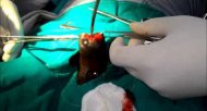
Abstracts: Transurethral resection of the bladder tumour (TURBT) is the standard surgical procedure for non-muscle invasive bladder cancer. We believe that all urologists should be trained in this procedure. This DVD provides an overview of TURBT with particular focus on basic skills, including basic surgical techniques such as the obturator nerve block. Important basic surgical skills required for complete TURBT in non-muscle invasive bladder cancer are: (i) resection of all visible tumors; (ii) resection of apparently normal mucosa on the border of the tumor; (iii) resection of the muscle layer at the base of the tumor until normal muscle fibers are visible; (iv) in applicable cases, random biopsy of apparently normal urothelium of the bladder wall and transurethral resection (TUR) biopsy of both sides of the prostatic urethra; and (v) when possible, after these procedures are completed, a different operating surgeon should inspect the bladder lumen to confirm that there are no remaining tumors. In particular, sampling resection should be implemented in apparently normal mucosa for approximately 1 cm around the tumor, and at the base of the tumor down to the superficial muscle layer. Resected specimens should be examined histopathologically in order to confirm the absence of malignant findings. Fundamental procedures for TURBT include both one-stage and two-stage resection. One-stage resection is used for relatively small tumors and involves a single procedure with simultaneous resection of both the tumor and the tissue at the tumor base down to the superficial muscle layer. In the two-stage resection, the first resection exposes the lower level of the mucosa and the second resection removes that lower mucosal layer in order to sample the superficial muscle layer for cancer staging. At the start of the resection, the loop is electrified before it makes contact with the mucosa. Delicate movements of the sheath should be used, along with delicate movement of the loop itself to adjust the depth of resection. The illustration of surgical techniques shows not only the basic techniques but also some points for caution during the resection. For actual resection, it is important to fully understand the properties of the tumor and to combine these techniques appropriately for each individual resection procedure. When resecting multiple tumors, the same basic resection techniques used for single tumors should be applied, and repeated as necessary.
This is a translated section of a video article originally published in Japanese as a DVD in the Audio-Visual Journal Vol.14 No.1. 2008 by The Japanese Urological Association. It has been modified with an English voice-over.